

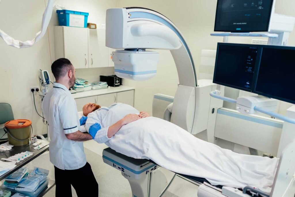
Medical imaging helps healthcare professionals examine internal body structures and understand different clinical conditions. While conventional X-rays produce still images, some medical procedures require continuous or repeated imaging to observe movement inside the body. This is where fluoroscopy becomes useful. It provides real-time X-ray imaging, allowing clinicians to observe anatomical structures and the movement of contrast materials during selected examinations and procedures.
What Is Fluoroscopy?
Fluoroscopy is a medical imaging technique that uses X-rays to produce moving images of the body’s internal structures. Unlike conventional radiography, which generally captures individual still images, fluoroscopy displays a sequence of images that allows movement to be observed.
It is used in several medical settings, including gastrointestinal examinations, orthopaedic procedures, vascular imaging and selected interventional procedures.
Depending on the examination, clinicians may use contrast materials to make certain organs, blood vessels or internal pathways easier to visualise.
How Does Fluoroscopy Work?
A fluoroscopy system generates X-rays that pass through the patient’s body and reach a digital detector. The detector captures the information, which is processed and displayed on a monitor as a real-time image sequence.
The imaging team can observe the movement of internal structures during the procedure and adjust the imaging equipment as required.
Modern systems may include image processing technologies designed to improve image presentation, manage noise and support radiation dose optimisation.
However, fluoroscopy involves ionising radiation, so examinations should be performed by qualified professionals using appropriate radiation protection practices.
Fluoroscopy vs Conventional Digital Radiography
Both fluoroscopy and digital radiography use X-rays, but they serve different imaging purposes.
Digital radiography (DR) generally captures individual images of anatomical structures. It is commonly used for chest, skeletal and other routine radiographic examinations.
Fluoroscopy, on the other hand, provides dynamic imaging. It allows clinicians to observe movement and changes over time rather than relying only on individual still images.
A conventional DR system is therefore generally suited to static radiographic examinations, while fluoroscopy is useful when real-time visual guidance or observation is required.
The choice between these techniques depends on the clinical question, examination requirements and the judgement of the treating medical team.
The Role of Image Processing in Fluoroscopy
Image processing plays an important role in dynamic imaging because fluoroscopic examinations involve sequences of images that must be displayed clearly and consistently.
Technologies designed for dynamic image processing can help manage image noise, brightness and contrast while maintaining the visual information needed during an examination.
Some advanced imaging platforms use specialised processing approaches for both fluoroscopy and rapid sequence imaging. These capabilities can support image quality and radiation dose optimisation.
The actual performance depends on the equipment, examination settings and clinical protocol.
How Fluoroscopy Differs from Mobile X-Ray Imaging
A mobile X-ray system is designed to bring radiographic equipment directly to patients, particularly when they cannot travel to a dedicated imaging room.
It is commonly used for bedside examinations in intensive care units, emergency departments and hospital wards.
Fluoroscopy serves a different purpose. Rather than focusing primarily on bedside mobility, it provides dynamic X-ray imaging for procedures that require observation of movement or real-time visual guidance.
Both technologies contribute to medical imaging, but their applications depend on different clinical requirements.
What Should Healthcare Facilities Consider?
When evaluating fluoroscopy equipment, healthcare facilities should consider image quality, dynamic imaging capabilities, radiation dose management, system flexibility and integration with existing radiology infrastructure.
Staff training, equipment maintenance, radiation protection and quality assurance are equally important.
Facilities should also evaluate how the system supports the specific examinations and procedures performed within their departments.
Understanding the Value of Dynamic Imaging
Fluoroscopy provides information that conventional still radiographs cannot always capture. By displaying movement in real time, it supports selected diagnostic examinations and image-guided procedures.
Understanding the differences between fluoroscopy, a DR system and a mobile X-ray system helps healthcare facilities identify the appropriate imaging technology for different clinical environments. Each has a distinct role in supporting medical imaging and patient care.